
Fig 5. A sequence of images of different frequencies (or times)
of the synchrotron emission from the same magnetic field configuration,
for a strong field model with B = 4 equivalent to a diffusion model with D = 0.0857.
As an example I show a sequence of images in Fig. 5 of the synchrotron emission from the same magnetic field configuration as before. The image changes quite dramatically as the spectrum ages. Prominent features at low frequency disappear entirely, to be replaced by new features. Most noticeable is the feature at the lower left in Fig. 5 which is prominent at low frequencies but is a definite hole in the high frequency image. The structures also become smaller and sharper as the spectrum ages.

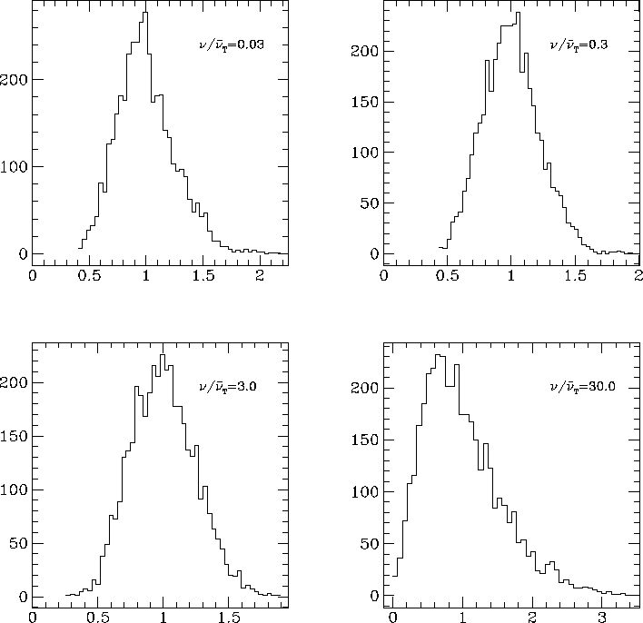
Fig 6. Histograms of pixel intensity for the same sequence of images as in Fig. 5. The shape of the histograms changes very little from a roughly bell-shaped distribution at low frequencies although a tail of high intensity points can just be seen at high frequency.
The shape of the intensity histogram (Fig. 6) changes slightly with frequency. At low frequencies the distribution of intensities is similar to a Maxwellian, and is roughly symmetric, so there are similar numbers of points above and below the median value. At higher frequencies a tail of high-intensity points starts to appear, although as in the intermediate model it is not as pronounced as the low field, high diffusion case.
The images are initially smooth and become more finely structured at higher frequencies. At high frequencies the image is mostly holes. At higher frequencies than shown here, this process continues. This leads to the appearance of thin, possibly filamentary structures. This is in accord with the image of Cygnus A obtained by Perley, Dreher & Cowan (1984), in which the filamentary structure is much more pronounced in the 6cm image than in the 20cm image and the filaments are most prominent in the oldest part of the lobes. Although these images were not at the same resolution, the results presented in this paper show that the increasing filamentation seen at high frequencies is not necessarily solely due to increased resolution. I discuss this point further in Section 6.
___________________________________ Peter Tribble, peter.tribble@gmail.com